Microscopic—and Huge
Bridges Powers Visualization of Whole Viruses at Atomic Level
An image can help human beings make connections. That’s why a scientist from Catholic University of America is using the large-memory nodes of PSC’s Bridges platform to put together some of the largest assemblies of proteins ever pictured. Thanks to Bridges’ powerful nodes with large amounts of memory, Victor Padilla-Sanchez has created images of whole viruses with atomic-level detail. These images will help engineer those viruses as vaccines or other medical therapies.
Why It’s Important
Math is central to science. But human beings are a very visual species. Sometimes, in addition to the math, we need a picture to really understand what’s going on. Scientists have had a lot of success visualizing all the atoms in biomolecules with precision, using computers. But some larger systems, such as whole viruses, have been beyond computers’ ability to visualize. In these cases, we rely on scientific artists, who use the best science available but still need to make interpretations. One famous example of this is the T4 bacteriophage, a virus that infects bacteria. Artists render it as something like a NASA lunar module. Something that looks like landing gear hangs at the bottom. A giant head at top contains the DNA payload that is so deadly to the bacteria it infects. But how accurate are these pictures? Artists may get the images mostly right. But they can’t know exactly how all the pieces fit together, which is what scientists need if they want to, for example, engineer T4 to kill the bacteria that cause diseases in humans.
“The goal of structural analysis is to investigate the interactions at atomic level between proteins. From there we can do many things. For example, we can … engineer the tail fibers of T4 to interact with the membrane proteins of other bacteria than E. coli. Or we can introduce [therapeutic] genetic material into other cells.”—Victor Padilla-Sanchez, Catholic University of America
Victor Padilla-Sanchez of Catholic University of America creates accurate computer-generated pictures of whole viruses. This is no small matter. Computer modeling has improved greatly over the years. But the state of the art today is to render pictures of protein assemblies that contain at most about 10 million of atoms. Visualizing whole viruses, on the other hand, involves structures that contain hundreds of millions of atoms. To achieve this, Padilla-Sanchez turned to the clever use of all the biochemical and microscope data available about a given virus and assembling the images in pieces, using the graphics processing unit (GPU) nodes and large memory of PSC’s Bridges supercomputer.
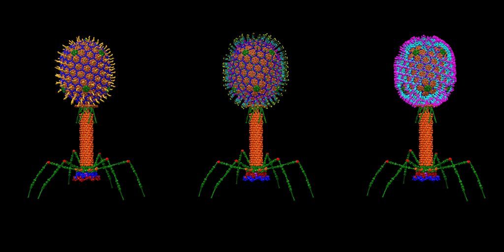
The T4 virus (left), modified to contain coat proteins from HIV, the AIDS virus (center), and the plague bacterium (right).
How PSC Helped
In previous work with computers at the Oak Ridge National Laboratory, Padilla-Sanchez assembled atom-by-atom images of T4 and HIV, the virus that causes AIDS. Using the University of California, San Francisco Chimera software, he used the computer to match the known 3D structures of the individual proteins in the virus to the fuzzy outlines of cryo-electron microscope images of the virus.
The images that he wanted to create in taking the work a step further would require juggling huge amounts of data. In order to render the images, the computer would have to access something like a terabyte of memory. Bridges, with 42 of its 3-terabyte large-memory (LM) nodes, had the power necessary for the job.
“The bacteriophage infection [image] is composed of approximately 210 million atoms. That figure was assembled with Bridges large memory using UCSF Chimera. I usually use one terabyte of RAM for my interactive sessions; the more memory I use, the more structures I can input in the software. Therefore, Bridges with its big memory—theoretically up to 12 TB RAM per session—gives a lot of computer power for this big-scale structural modeling.”—Victor Padilla-Sanchez, Catholic University of America
Two of Padilla-Sanchez’s new findings with Bridges LM included an image of three T4 viruses. One has its natural coat proteins. The other two have been virtually engineered to display the coat proteins from HIV-1 or the plague bacterium. These two could be vaccine candidates for AIDS or the plague. Another Bridges image is a time series showing a T4 phage “landing” on the surface of a bacterium, much like a lunar lander coming down. Each of these images will be useful in engineering T4’s behavior to serve medical purposes. Padilla-Sanchez reported these results in the proceedings of the Institute of Electrical and Electronics Engineers’ VIS 2019 conference in Vancouver in October.
Next steps will include imaging larger systems as well as T4 engineered to serve as a vaccine. Padilla-Sanchez plans to expand his software to include Maestro—software that handles better electrostatics visualization and has many other features that Chimera does not have—and ChimeraX, a next-generation version of Chimera with improved functionality. In the future, with more powerful computers, such predicted structures may be used to run molecular dynamics simulations studying the movements of proteins within whole viruses rather than the static pictures now possible. He plans to investigate the movements of T4 to start with, and then move on to other organisms.