
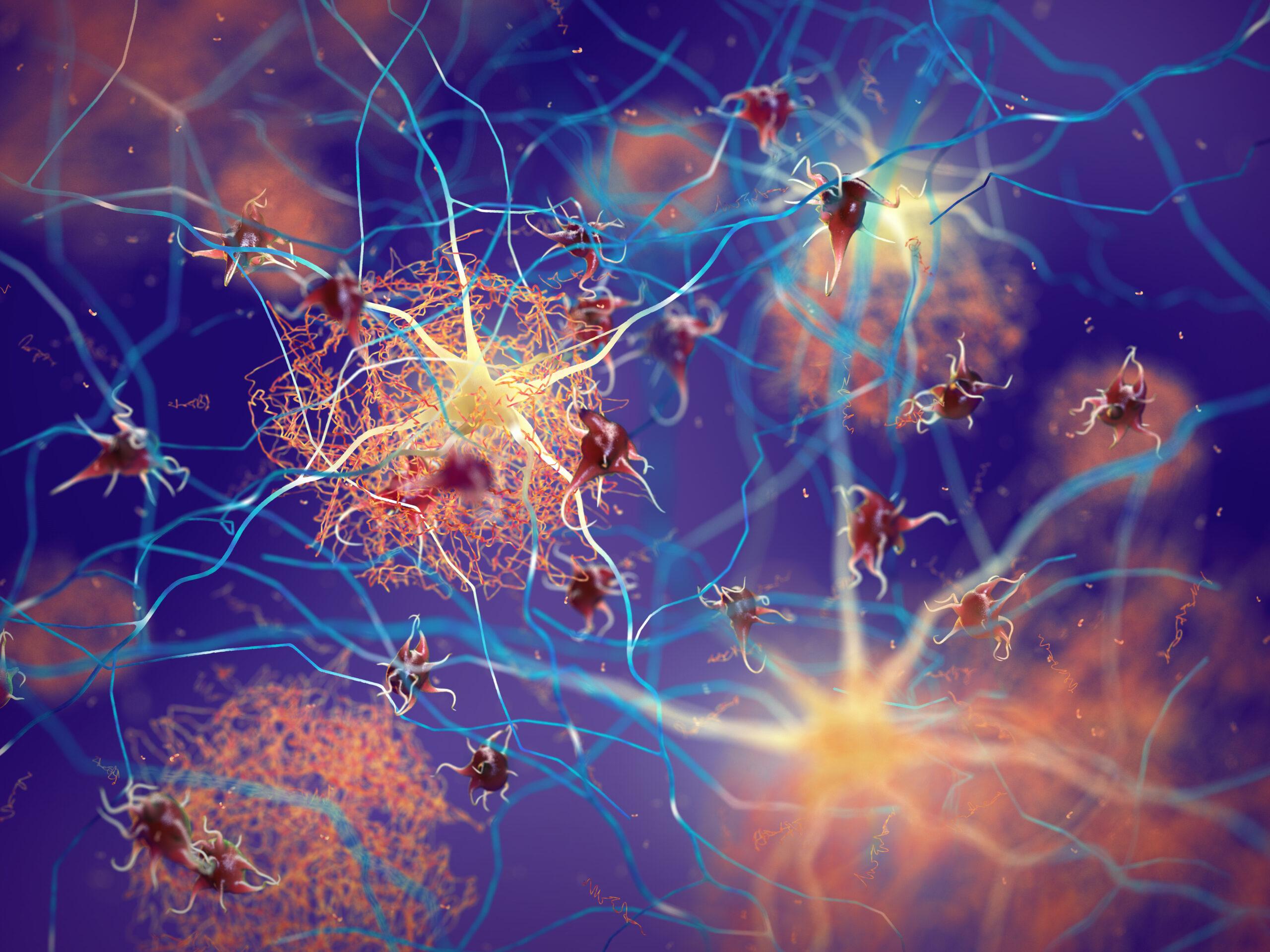
Amyloid plaques forming between neurons. Beta-amyloid protein disrupting nerve cells’ function in a brain with Alzheimer’s disease.
Bridges-2 Simulations Point to Process of Fibril Formation, Offering Clues to Combatting ALS, Alzheimer’s, Parkinson’s
Fibril forming proteins, which stick together to make protein chains that gum up a cell’s works, underlie many degenerative diseases, like Alzheimer’s, Parkinson’s, and ALS (Lou Gehrig’s disease). Attempts to prevent disease processes with anti-fibril drugs have been disappointing, though. A team led from the University of Texas at Austin (UT Austin) used PSC’s flagship Bridges-2 supercomputer in a series of simulations that uncovered the process of how an ALS-associated protein triggers fibrils. The results offer clues as to how fibril formation happens, and possibly how it could be better prevented.
WHY IT’S IMPORTANT
While proteins often float in the liquid of the cell’s cytoplasm, they can sometimes form solid structures. Some of these structures, like the fibers in muscle cells, are part of healthy cell activity. But they can also lead to human diseases like Alzheimer’s, Parkinson’s, and ALS. In those maladies, proteins that normally carry out their functions in the cell’s liquid phase suddenly begin to aggregate. They form fibrils, lengthening ropes of protein that accumulate and begin to destroy the cell that contains them. This can start deterioration of memory (Alzheimer’s), brain control of muscles (Parkinson’s), and the nerve cells that connect with muscles (ALS). Many attempts have been made to short-circuit these diseases with drugs that prevent fibril formation, particularly Alzheimer’s. But so far, the results have been disappointing.
“There was a paper in Cell some few years ago … which said that, upon phase separation … [fibril-forming proteins are] more solid-like. And there were several papers that said the opposite. Even in the particular problem of FUS, there’s a … disagreement within experiments. So, it’s not fully settled, as far as I can tell. And that’s one of the reasons we have thought about … FUS, and we want to understand the molecular origins of what’s going on.”
— Dave Thirumalai, UT Austin
A collaboration of scientists at UT Austin, Georgetown University, and the Universidad Nacional Autónoma de México, led by Dave Thirumalai at Austin, wanted to understand the process of fibril formation and such phase transitions in proteins at the fundamental level. This basic science knowledge, they reasoned, could give drug researchers the tools needed to improve their batting average against malignant fibril formation. A major tool in the team’s investigations has been a series of protein simulations using PSC’s flagship Bridges-2, available to them through the NSF’s ACCESS network of supercomputers, in which PSC is a leading member.
HOW PSC HELPED
The UT-led group focused on the Fused in Sarcoma protein’s low-complexity domain (FUS-LC). An important actor in sarcoma cancers as well as the development of ALS, FUS consists of two sections. One part has a “normal” protein structure, with varied amino acid components that interact to fold into a set structure. The LC part, though, has much less varied amino acids, and so is intrinsically disordered. That is, it exists as a wiggly chain of amino acids without a set folding pattern. Scientists had isolated the LC domain of FUS to create FUS-LC, a wiggly chain that has no set structure.
Until it does.
Like the full FUS protein, FUS-LC can eventually fold into a form that aggregates to build fibrils. Because it’s smaller than full FUS and doesn’t include the more complex parts of the protein, FUS-LC’s movements and fibril formation are easier to simulate and study. Using Bridges-2, Thirumalai’s team studied this structure to pick apart the exact sequence of how FUS-LC takes its toxic, fibril-friendly form.
Bridges-2 was ideal for the work. Offering 47 graphics processing unit nodes, with a total of 384 GPU processors, Bridges-2 allowed the team to simulate the complex movements of multiple FUS-LC molecules as they folded and interacted with each other. The speed with which Bridges-2 could carry out these sims also helped the scientists carry out the many repeated runs they needed for significant results.
“Currently, Bridges-2 is really helping us, because we have developed these simplified models that allow us to simulate 500 [protein] chains, plus the monomers … reasonably well … We can’t declare victory here now. We have to think about this. But in the absence of the GPUs … It would take several years to do what we did in this PNAS paper, for example.”
— Dave Thirumalai, UT Austin
The simulations revealed that FUS-LC follows a relatively set pattern in folding and aggregating. The three parts of FUS-LC that first fold, called core-1, -2, and -3, did so in a regular order. Core-3, which has the least stable fold, forms first. That’s because, as the most disordered, its transition from wiggly to folded is the smallest change. Core-1, the most stable when folded, forms last, as it has to overcome a larger transition.
This result extends Ostwald’s rule of stages, originally formulated for crystalline systems, to biomolecules. This shows that staged transformations similar to those seen in crystals also govern fibril formation in protein assemblies. Moreover, core-3’s folding helps to trigger the folding of core-2 and core-1. This causes a chain reaction that makes FUS-LC begin to aggregate with other FUS-LC molecules, another sign of Ostwald ripening. The scientists reported their results in the Proceedings of the National Academy of Sciences USA in January 2026.
The team’s findings point the way to how other fibril-forming proteins may begin to aggregate. One such protein is TAR DNA-binding protein 43 (TDP-43), a similar protein to FUS that also plays a role in ALS, as well as several types of dementia including Alzheimer’s. On a more basic level, the results may help scientists understand better how other fibril-forming proteins aggregate, as well as the still-mysterious roles these proteins must play, when they don’t aggregate, in healthy cell function.